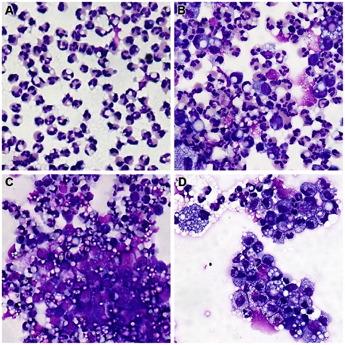
Figure 3. Microscopic examination of milk cells recruited at different times following intramammary infusion with ovalbumin.
Milk cells from quarters infused with ovalbumin were cytocentrifuged on glass slides and stained with May-Grünwald-Giemsa. Slides representative of each sampling time are shown. A) 12 hpi; B) 24 hpi; C) 48 hpi; D) 96 hpi;

