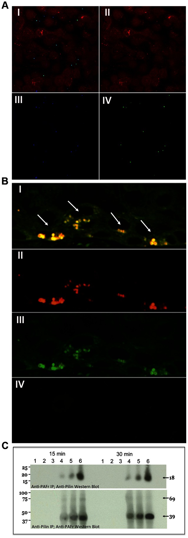
Figure 4. N. meningitidis C311#3 pilin-linked ChoP mediates adherence to the PAFr on human airway epithelial cells.

(A) Confocal microscopy demonstrates that nearly 100% of N. meningitidis C311#3gfp co-localize with the PAFr on 16HBE14 cells at 15 minutes post-infection. Panel (I) shows the merged image of red, blue, and green channels. Panel: (II) Red channel - Cell Tracker-stained 16HBE14 cells, (III) Blue channel - PAFr, (anti-PAF rabbit sera+Alexa 647 goat anti-rabbit), (IV) Green channel - GFP-expressing C311#3. (B) Co-localization of C311#3 WT (red) with the PAFr (green) is readily visible by confocal microscopy as a yellow fluorescence (denoted by arrows) following a 30 minute infection of human bronchial tissue. Panel (I) shows the merged image of the red and green channels. Panel: (II) Red channel - N. meningitidis immunolabeled with antibody pS-20 and a rhodamine-conjugated secondary antibody, (III) Green channel - the PAFr immunolabeled with primary antibody H-300 and a FITC-conjugated secondary antibody (IV) Control in which both primary antibodies have been omitted. (C) IP using anti-pilin (top panel) or anti-PAFr (bottom panel) antibodies demonstrates a direct interaction occurs between the PAFr and pilin at 15 and 30 minutes post-challenge of 16HBE14 cells. Lanes: (1) the primary antibody was omitted from the capture step, (2) the secondary, agarose-conjugated, antibody was omitted from the assay, (3) uninfected 16HBE14 cells, infections were performed using (4) strain C31126A (trisaccharide, no ChoP), (5) strain C311pglA (disaccharide, ChoP+), and (6) WT C311#3 (trisaccharide, ChoP+). These data show that both ChoP and the pilin-linked glycan are required for efficient binding to the PAFr on human bronchial cells.
