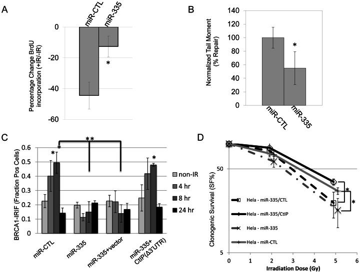
Figure 4. DDR defects induced by miR-335 overexpression in HeLa cells.
(A) RDS (radioresistant DNA synthesis): miR-CTL and miR-335 overexpressing HeLa cells were treated with or without 10 Gy IR and S-phase DNA synthesis was labeled with BrdU. The percent change of BrdU incorporation (+IR/−IR) was summarized from three independent experiments. MiR-CTL overexpressing HeLa cells showed a reduction (42%) in the percentage of BrdU incorporation whereas miR-335 overexpressing HeLa cells showed less of a reduction (16%). The * indicates p<0.05. (B) NCA (neutral comet assay): quantification of DNA repair with the NCA indicates reduced DNA repair at 5 hours after 15 Gy in HeLa cells overexpressing miR-335. Three independent experiments were performed; the * indicates p<0.05. (C) BRCA1 foci: immunofluorescent staining of HeLa cells after 12 Gy IR with BRCA1 showed a foci-forming defect in miR-335 overexpressing cells (bars 7 and 11). This response was reversed and corrected in miR-335 overexpressing cells co-transfected with CtIP lacking the 3′UTR (Δ3′UTR) (bar 15). The * indicates intra-sample Student's t-test (p<0.05) comparing the indicated bar with the non-IR condition. ** indicates inter-sample Student's t-test (p<0.05) comparing the 8 hour time points between samples. (D) CSA (clonogenic survival assay): reduced colony survival in miR-335 overexpressing HeLa cells post-IR. The survival fraction was significantly improved when CtIP (Δ3′UTR) was added back to miR-335 overexpressing HeLa cells. The radiation dose used for all samples is 0, 2, and 5 Gy. The x-axis has been offset for each pair of data points to make viewing the data and error bars easier. The * indicates p<0.05.

