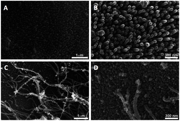
Figure 2. Surface characteristics of Schistosoma mansoni and S. eggshells.
Panels A and B show the surface ofjaponicum a S. mansoni egg imaged with high resolution scanning electron microscopy illustrating that the surface is completely covered with filaments or microspines. Figures C and D show similar observations of S. japonicum. The microspines on the surface of S. japonicum are shorter and the surface is covered with an additional filamentous matrix.

