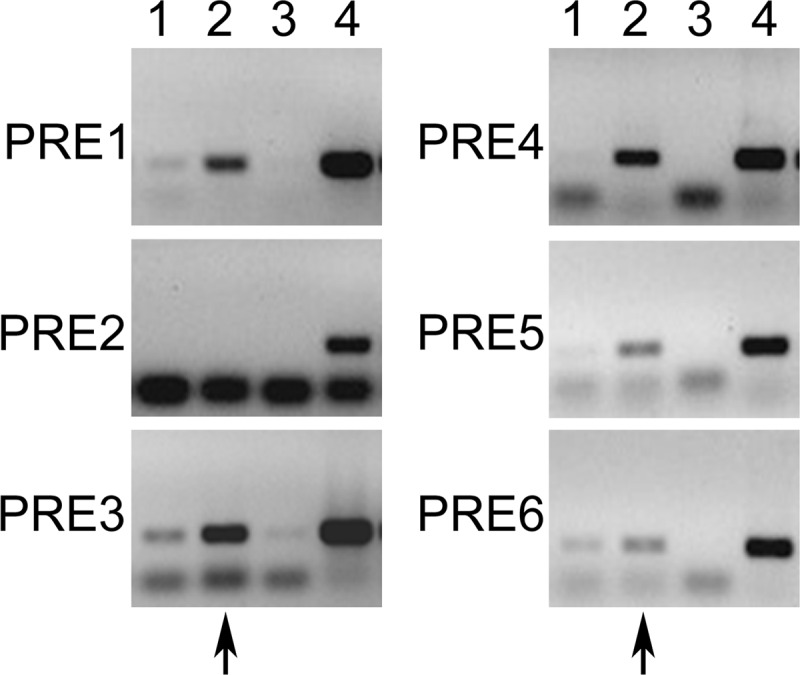
Figure 4.

ChIP PCR results from the different PRE sites present in the −5.7;−4.2-kb region. PRE1 to -6 were tested individually, and immunoprecipitation was carried out with the anti-PRB antibody. Cells were transfected with hPRB and treated with the carrier (lane 1) or progesterone (lanes 2, 3, and 4). Lane 3 is a negative control with an unrelated antibody or IgG, and lane 4 is an input control. The observed lower bands are the oligonucleotide dimers. The arrows indicate the lane where a positive PCR amplification is expected.
