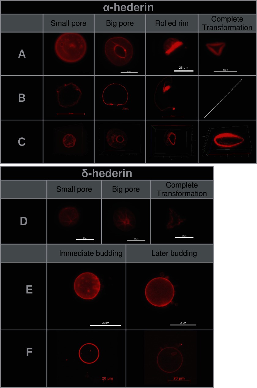
FIGURE 10.
Stages of pore formation (A–D) and budding (E and F) of GUVs composed of DMPC/Chol (3:1) incubated with 40 μm of α-hederin (A–C) or δ-hederin (D–F). A, D, and E, fluorescence microscopy images of vesicles; one bar represents 25 μm. B and F, confocal microscopy, cross-section of the middle of one vesicle, one bar represents 20 μm. C, confocal microscopy, three-dimensional view of all cross-sections taken from one vesicle.

