FIGURE 7.
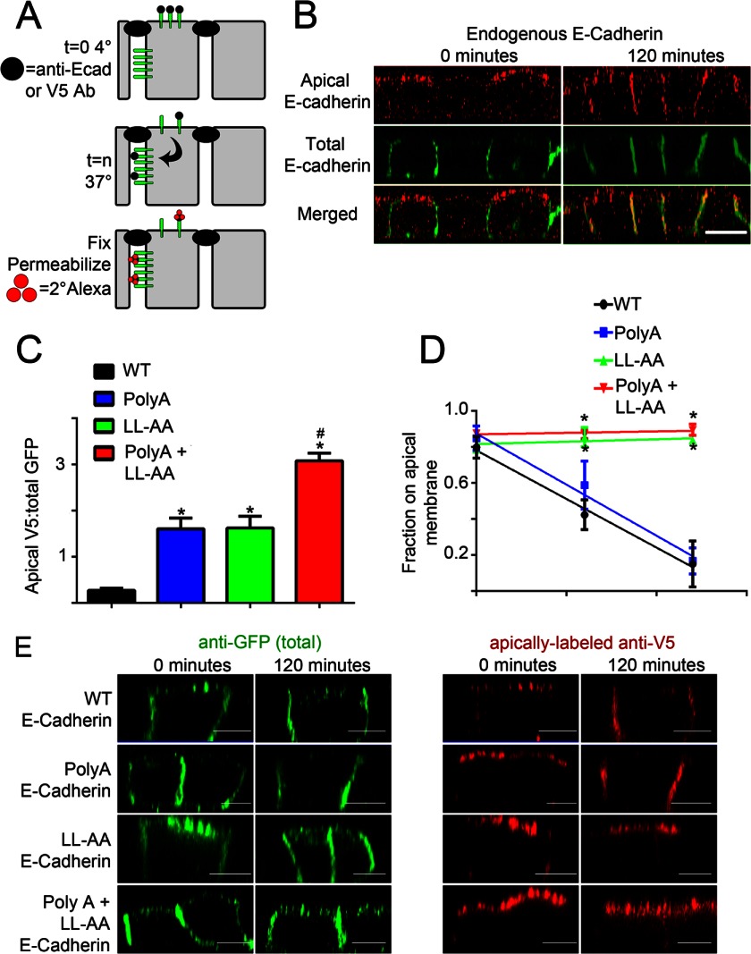
Dileucine/alanine mutation abolishes editing of apically mislocalized E-cadherin in MDCK cells. A, a model of apical E-cadherin internalization assay is shown. Confluent, filter-grown MDCK cells were transfected with E-cadherin containing an extracellular V5 epitope and a cytoplasmic GFP epitope. Cells were then labeled on the apical surface with primary antibody (black circle) on ice for 1 h followed by extensive washing. Cells were warmed to 37 °C for the indicated times to allow trafficking of E-cadherin from the apical membrane. Cells were then fixed, permeabilized, and labeled with secondary antibodies tagged with Alexa568 (red circles). B, shown are representative XZ projections of MDCK cells stained with antibodies to endogenous E-cadherin. Total E-cadherin is marked with mouse anti-E-cadherin antibodies (green). Apically labeled antibody to the extracellular epitope of E-cadherin (rat anti-E-cadherin) is shown in red. Bars represent 10 μm. C, shown is quantification of the amount of apical V5 labeling of V5-E-cadherin and mutants normalized to total GFP expression. *, p < 0.05 compared with wild-type. #, p < 0.05 compared with both Poly(A) and LL-AA mutants (one way ANOVA followed by Tukey post hoc test; n = 5 cells per condition). D, shown is the quantification of the fraction of apically labeled E-cadherin remaining on the apical surface. Mean pixel intensity of a three-dimensional region of interest containing the apical membrane was quantified and compared with a three-dimensional region of interest containing the lateral membrane. *, p < 0.05 compared with both wild-type and Poly(A) E-cadherin (two-way ANOVA followed by Tukey post hoc test, n = 5 cells for point). E, shown are representative XZ projections of MDCK cells transfected with wild-type (top), Poly(A) (2nd row), LL-AA (3rd row), or Poly(A) + LL-AA mutant (bottom) E-cadherin warmed to 37 °C for 0 min (left) or 120 min (right). Total E-cadherin-GFP is marked with anti-GFP antibodies and is shown in green (left). Apically labeled anti-V5 to the extracellular epitope of E-cadherin is shown in red (right). Bars represent 5 μm.

