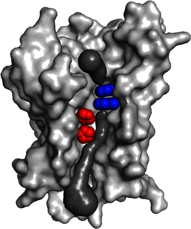
FIGURE 1.
Substrate conduction path of E. coli AmtB. The AmtB monomer as viewed from the membrane, with the periplasmic surface at the top and cytoplasmic surface at the bottom. Forward facing transmembrane helices have been removed to reveal the substrate transport path (modeled in dark gray). Components of the conserved twin-histidine element (His168 on the top and His318 on the bottom) are highlighted in red. A constriction composed of two conserved and presumably mobile phenylalanine residues (Phe107 on the top and Phe215 on the bottom) that separates the periplasmic vestibule of the transport pathway from the conduction pore is shown in blue. Whether ammonium crosses the phenyl ring constriction and traverses the conduction pore as NH3 or NH4+ remains a point of debate. The AmtB model was created from Protein Data Bank entry 2NS1 (26) using PyMOL, and the substrate conduction pathway was visualized using the program CAVER (27).

