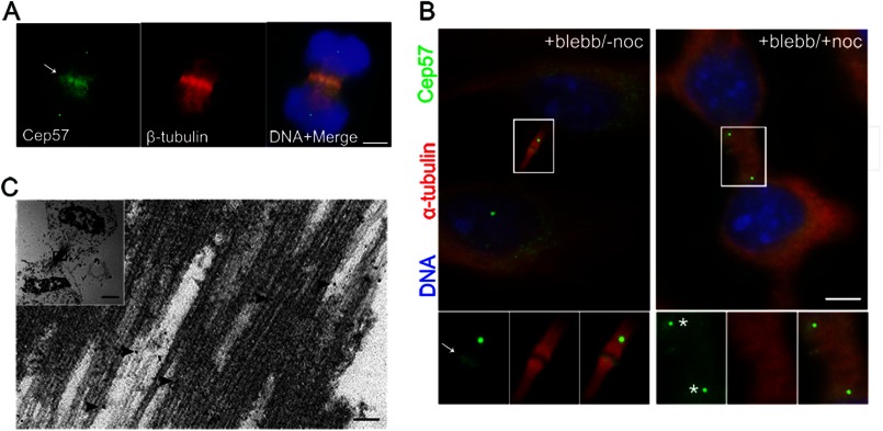
FIGURE 1.
Cep57 is a central spindle and midbody component. A, indirect immunofluorescence of anaphase cells with anti-Cep57 (green) and anti-β-tubulin (red) antibodies. DNA was labeled with DAPI (blue). The arrow indicates Cep57 in the midzone. Scale bar = 5 μm. B, HeLa cells were treated with 100 μm blebbistatin (blebb) for 2 h. After washout of blebbistatin, cells were subjected to fresh 37 °C culture medium or to 10 μg/ml nocodazole (noc) at 0 °C for 1 h. Shown is the indirect immunofluorescence of cells with anti-Cep57 (green) and anti-α-tubulin (red) antibodies. DNA was labeled with DAPI (blue). The arrow indicates Cep57 at the midbody. The asterisks indicates Cep57 at the centrosome. Scale bar = 5 μm. C, immunoelectron microscopy images of Cep57 localization within the midbody of a HeLa cell. Arrowheads indicates antibody-conjugated gold particles. The inset shows a lower magnification image of the cell undergoing cytokinesis. Scale bars = 100 nm and 2 μm (inset).

