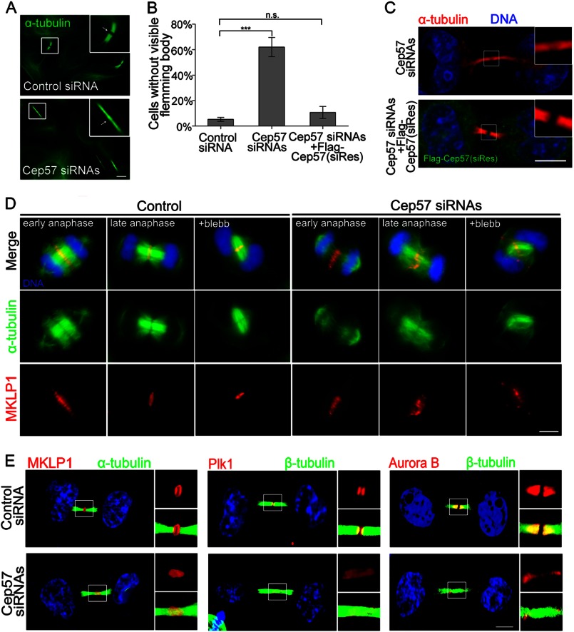
FIGURE 3.
Depletion of Cep57 perturbs the microtubule organization of the central spindle and the localization of the midbody components MKLP1, Plk1, and Aurora B. A and B, HeLa cells transfected with control siRNA, Cep57 siRNA, or Cep57 siRNA and FLAG-tagged siRNA-resistant Cep57 (siRes) were treated with 2.5 mm thymidine for 24 h to G1/S phase and then released for 12 h to telophase. The cells were stained with anti-α-tubulin antibody (A; green). Scale bar = 5 μm. B shows the increased percentage of Cep57-depleted cells with no split site (A, arrow in lower panel) at the midbody region. A minimum of 100 cells were counted per sample in three independent experiments. Error bars represent ± S.E. ***, p < 0.001; n.s., not statistically significant. C, immunofluorescence images of HeLa cells treated with the indicated siRNAs for 60 h. The cells were stained with anti-α-tubulin (red) and anti-FLAG (green) antibodies. DNA was labeled with DAPI (blue). Scale bar = 5 μm. D, immunofluorescence images showing that depletion of Cep57 disrupts central spindle microtubule organization and MKLP1 localization. Images are shown of control and Cep57-depleted cells in anaphase with or without blebbistatin (blebb) treatment. The assembly state of the anaphase central spindle is shown by α-tubulin (green), MKLP1 (red), and DNA (blue). Scale bar = 5 μm. E, immunofluorescence images of Cep57-depleted and control cells. The cells at telophase were stained for MKLP1 (red), Plk1 (red), Aurora B (red), tubulin (green), and DNA (blue). Scale bar = 5 μm.

