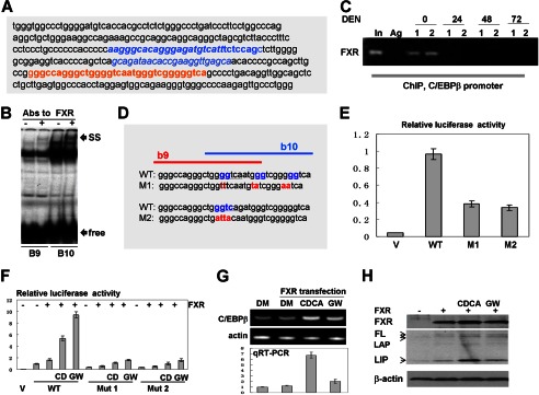
FIGURE 6.

FXR up-regulates C/EBPβ through direct binding to the C/EBPβ promoter. A, nucleotide sequence of the C/EBPβ promoter containing FXR binding sites. Overlapping b9 and b10 FXR consensuses are shown in red. B, EMSA with probes covering FXR binding sites b9 and b10. Abs to FXR were incorporated in the binding reactions. Positions of supershift (SS) and free probes (free) are indicated by arrows. C, FXR binds to the C/EBPβ promoter in quiescent livers but is removed from the promoter in livers at early steps of DEN-mediated cancer. A ChIP assay was performed with chromatin solutions from quiescent livers (0) and livers at 24, 48, and 72 h after DEN injections. Data with two animals for each time point are shown. In, 1/100 input; Ag, agarose beads with IgG from preimmune serum. D, incorporation of mutations in the FXR binding sites within the C/EBPβ promoter. The incorporated mutations are shown in red. E, the mutations of the FXR binding site within the C/EBPβ promoter reduce the activity of the promoter. The Luc-WT and M1 and M2 mutant C/EBPβ promoters were transfected in Hep3B2 cells. The luciferase activity was examined as described above, and it is shown as a ratio to Renilla control. V, vector. F, FXR activates the C/EBPβ promoter through FXR binding sites. CD, CDCA; GW, GW4064. WT and mutant promoters (M1 and M2) were cotransfected with FXR into Hep3B2 cells. Cells were treated with the FXR ligands CDCA and WG4064. DM, dimethyl sulfoxide. G, activation of FXR by ligands increases expression of endogenous C/EBPβ mRNA. The top panel shows a typical picture of a regular RT-PCR assay. β-actin was used as the control. The bottom panel shows the results of quantitative RT-PCR (qRT-PCR) analysis of C/EBPβ mRNA. H, Western blotting shows activation of the endogenous C/EBPβ by FXR. The upper panel shows Western blotting with Abs to FXR. The C/EBPβ filter was reprobed with Abs to β-actin (bottom panel). Positions of the full-length (FL), LAP, and LIP isoforms of C/EBPβ are shown by arrows.
