FIGURE 5.
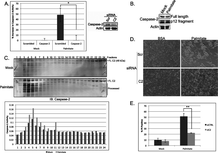
Caspase-2 plays a central role in mediating lipoapoptosis in 293T cells. A, 293T cells were transfected with 50 nm scrambled (Scr) or caspase-2 (C2)-targeted siRNA. At 48 h post-transfection, cells were mock-treated with BSA or treated with 1 mm palmitate for 18 h, and caspase-2 activity was assessed using the BioVision caspase-2 colorimetric assay kit. Knockdown efficiency was determined by immunoblot. The results represent the means ± S.E. (error bars) for percent increase in activity compared with control siRNA-, mock-treated samples for three independent experiments. Statistical significance was determined using a two-tailed Student's t test. *, p < 0.05 versus control. B, 293T cells were treated with BSA or palmitate for 24 h and immunoblotted for caspase-2 and actin. The top caspase-2 panel is a short exposure (to show changes in full-length caspase-2 levels), and the bottom caspase-2 panel is a long exposure (to reveal the p12 fragment). C, 293T cells were treated with BSA or palmitate for 18 h before lysis by Dounce homogenization in hypotonic lysis buffer. Cell lysates were separated on a Superdex 200 column, and relevant fractions were immunoblotted (IB) for caspase-2. Full-length caspase-2 (FL C2) and processed caspase-2 are indicated. Fractions were also tested for caspase-2 activity by assessing cleavage of the caspase-2 model substrate, VDVAD-pNA, as part of the BioVision caspase-2 colorimetric assay kit. D, 293T cells were treated as in A, and at 24 h post-treatment, phase-contrast images were captured on an EVOS FL digital inverted microscope. E, 293T cells were treated as in A, and cell death was measured 24 h after palmitate treatment by propidium iodide staining and flow cytometric analysis. The results represent the means ± S.E. (error bars) for four independent experiments. Statistical significance was determined using a two-tailed Student's t test. **, p < 0.01 versus control. siCTRL, control siRNA; siC2, caspase-2 siRNA.

