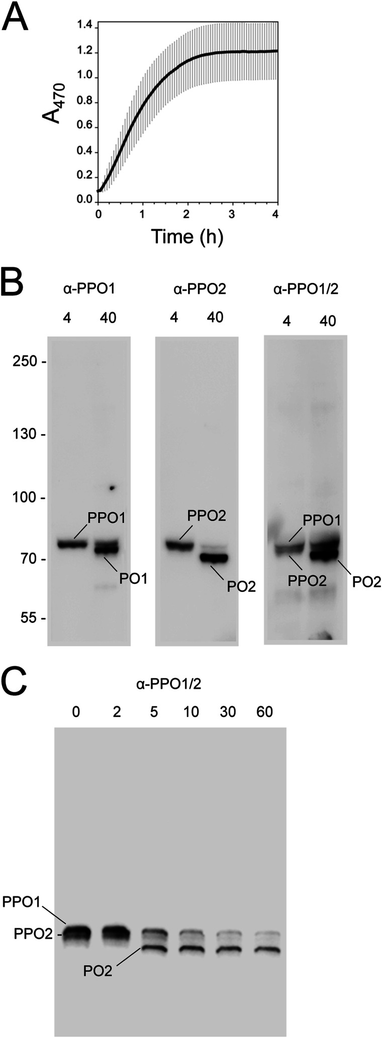
FIGURE 1.
The PO cascade in B. mori is activated by wounding. A, melanization rate of B. mori plasma. Freshly prepared plasma from three fifth instar larvae (20 μl each) was diluted in PBS (80 μl), plated, and monitored for melanization activity 0–4 h ± S.D. post-collection from a wound site. Activity was measured by monitoring formation of dopachrome or dopaminechrome (melanization intermediates) at A470. B, plasma was collected as outlined in A, followed by the addition of sample buffer at 4 or 40 min. After separation on 7.5% continuous SDS-polyacrylamide gels under reducing conditions, samples were immunoblotted using antibodies for PPO1 (α-PPO1), PPO2 (α-PPO2), or a cross-reacting antibody that recognizes both PPOs (α-PPO1/2). Bands corresponding to the predicted sizes of PPO1/PO1 and PPO2/PO2 are indicated. Note that the affinity of α-PPO1/2 for PO2 is stronger than for PO1. C, immunoblot prepared as described in B but with sample buffer added at 0–60 min post-collection and probed using α-PPO1/2. Bands corresponding to PPO1/PO1 and PPO2/PO2 are indicated. Molecular mass markers in B are indicated on the left.

