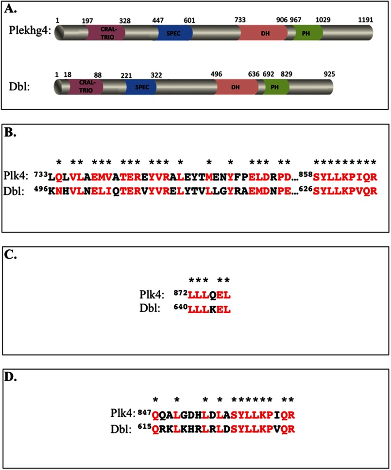
FIGURE 2.
Sequence signatures of Plekhg4 and Dbl. A, domain structure of the two proteins inferred using sequence prediction algorithm (Expasy). Domain size and limits were manually drawn to scale. B–D, sequence homology between Plekhg4 and Dbl. Shown are sequence alignments between the DH domains of the proteins (B), residues that mediate GTPase binding (C), and the residues that dictate GTPase selectivity (D). Identical and highly similar residues are shown in red and denoted by asterisks.

