FIGURE 7.
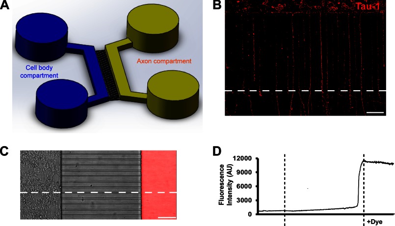
Microfluidic chambers direct axon growth of hippocampal neurons and fluidically isolate axons. A, schematic of a microfluidic chamber. B, image of neurons (DIV5) cultured within the microfluidic chamber, stained for tau-1 (red). Scale bar, 100 μm. C, Alexa Fluor 594 dye (20 μg/ml) was added to the axon compartment at DIV4. The image is an overlay of the phase contrast and fluorescence images after 24 h of treatment. Scale bar, 100 μm. D, fluorescence intensity change trend along the horizontal dotted line through the entire image shown in C.

