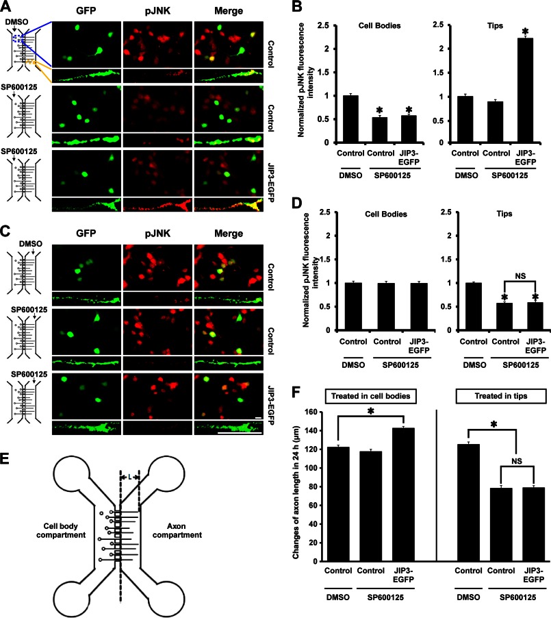
FIGURE 8.
JIP3 enhances axon elongation by locally activating JNK at axon tips. A and C, hippocampal neurons transfected with empty vector or JIP3-EGFP construct were cultured in the microfluidic chambers for 4 days. Then the JNK inhibitor SP600125 (10 μm) was applied to the cell body compartment (A) or the axon compartment (C) for 24 h. Cells were stained with anti-GFP (green) and anti-pJNK (red) antibodies. Images of cell bodies and axon tips are shown. Scale bar, 20 μm. B and D, quantification of results from A and C. The graphical data shown on the left of B and D represent the relative fluorescence intensity of pJNK in the cell bodies, and those on the right represent that at axon tips (n = 3, *, p < 0.05, versus the control + DMSO group; NS, no significance; Student's t test). E, schematic for calculation of the axon length in a microfluidic chamber; “L” represents the axon length of interest. F, statistical results for the changes in “L” in E over 24 h, during which the cell bodies or axon tips were treated with DMSO or SP600125 (n = 3, *, p < 0.05, versus the control + DMSO group; NS, no significance; Student's t test).

