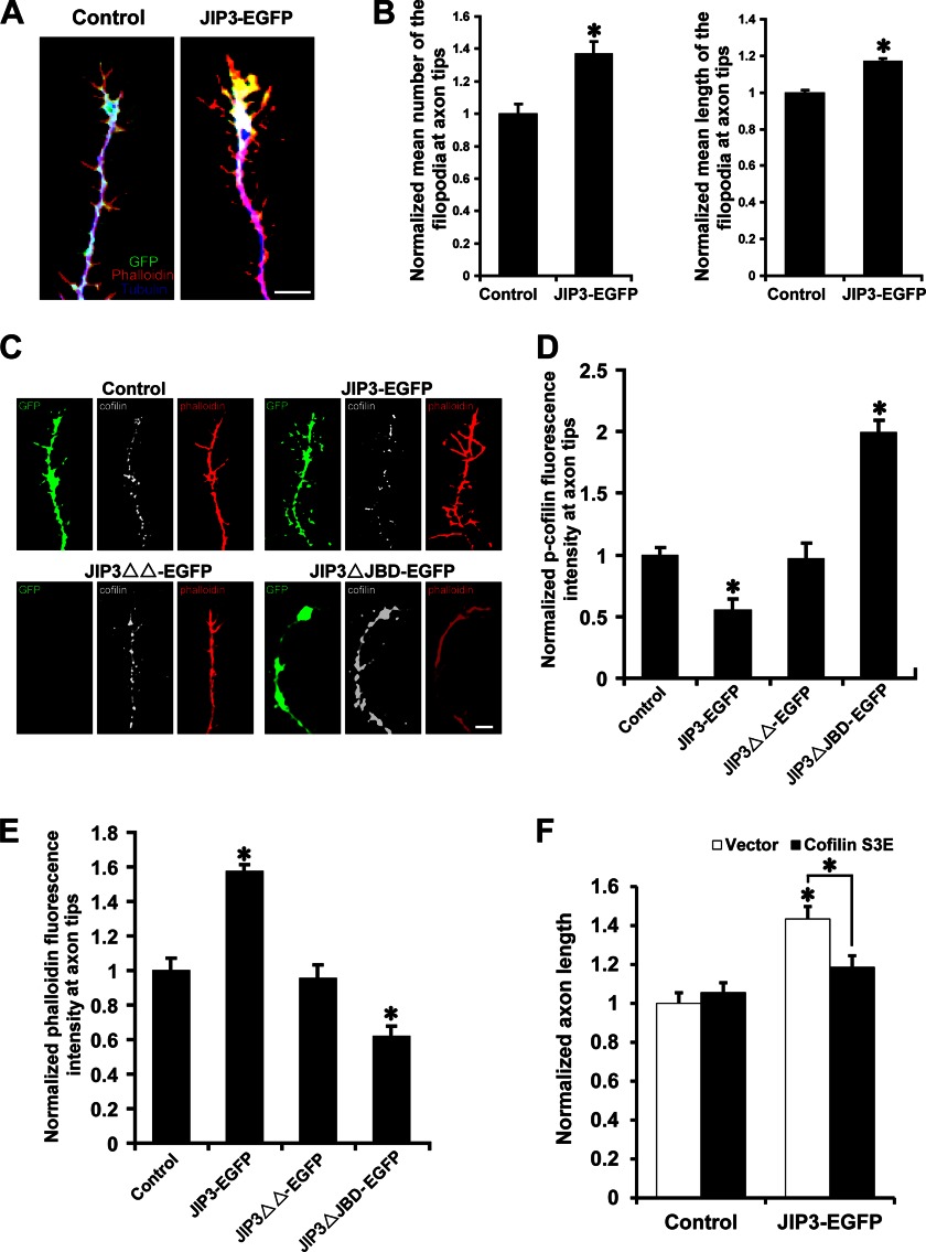
FIGURE 9.
JIP3 regulates actin dynamics at axon tips through the JNK-cofilin pathway. A, representative images of axon tips from neurons (DIV5) transfected with empty vector or JIP3-EGFP. Cells were stained with anti-GFP (green) and anti-α-tubulin (blue) antibodies. F-actin (red) was labeled with Alexa Fluor 594-conjugated phalloidin. Scale bar, 5 μm. B, quantitation of the relative number and length of filopodia at axon tips shown in A (n = 3, *, p < 0.05, versus the control group; Student's t test). C, representative images of axon tips from neurons (DIV5) transfected with the indicated constructs. Cells were stained with anti-GFP (green) and anti-p-cofilin (gray) antibodies. F-actin (red) was labeled with Alexa Fluor 594-conjugated phalloidin. Scale bar, 5 μm. D and E, quantitation of the staining intensities of p-cofilin (D) and F-actin (E) at axon tips shown in C (n = 3, *, p < 0.05, versus the control group; Student's t test). F, quantitation of the axon length in neurons co-transfected with empty vector or JIP3-EGFP and control or cofilin S3E constructs (n = 3, *, p < 0.05, versus the control + vector group; one-way analysis of variance).

