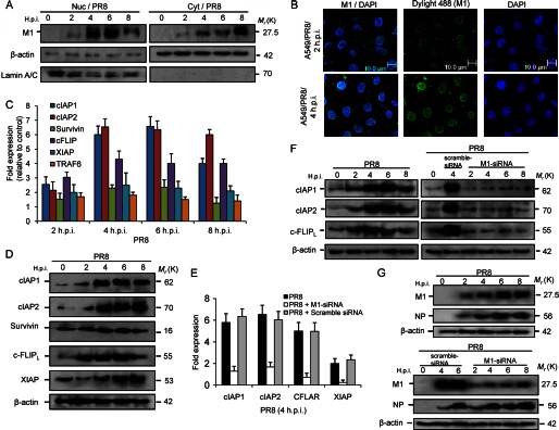
FIGURE 1.

M1 up-regulates survival gene transcription in PR8-infected A549 cells. A, expression of M1 protein was observed after whole cell lysates were fractionated to nuclear (Nuc) and cytoplasmic (Cyt) fractions and immunoblotted from control and PR8-infected (1 m.o.i.) cells for the given time points (0, 2, 4, 6, and 8 h.p.i.). Lamin A/C is a loading control for the nuclear fraction, and β-actin is a loading control for the cytoplasmic fraction. B, confocal imaging of A549 cells after PR8 infection (1 m.o.i.) at early infection periods (2 and 4 h.p.i.) was performed to analyze cellular (nuclear) localization of M1 protein during infection. DyLight 488 (green) indicates M1 protein, and DAPI (blue) indicates the nucleus. C, mRNA levels of the RelB candidate target genes including cIAP1, cIAP2, survivin, cFLIP, XIAP, and TRAF6 were analyzed in A549 cells after the cells were infected with influenza A/PR8 virus (1 m.o.i) for the given time points. RNA (1 μg) was converted to cDNA by Superscript II, amplified by Q-PCR (40 cycles), and then quantified by SYBR Green fluorescence. GAPDH was used as a reference gene. Data are presented (for PR8-infected cells) as -fold change (based on 2ΔΔCt values) relative to non-infected control cells (mean ± S.D.; n = 3). D, A549 cells were infected with PR8 strain (1 m.o.i.) for the given time points (0, 2, 4, 6, and 8 h.p.i.), and the whole cell lysates of the infected and mock-infected (control) cells were subjected to immunoblotting to analyze protein levels of cIAP1, cIAP2, survivin, cFLIP, and XIAP. β-Actin served as an internal loading control. E, M1 siRNA (60 nmol) along with scrambled siRNA (60 nmol; for siRNA control) was transfected to A549 cells prior to PR8 infection (1 m.o.i), and real time analysis of cIAP1, cIAP2, cFLIP, and XIAP transcripts was performed by Q-PCR. GAPDH was used as a reference gene. Data are presented as -fold change (based on 2ΔΔCt values) relative to non-infected control cells (mean ± S.D.; n = 3). F, protein levels of antiapoptotic genes in only PR8-infected (1 m.o.i.), scrambled siRNA (60 nmol)-treated PR8-infected (1 m.o.i.), and M1 siRNA (60 nmol)-treated PR8-infected (1 m.o.i.) A549 cells were analyzed by immunoblotting using β-actin as an internal loading control for the given time points (0, 2, 4, 6, and 8 h.p.i.). G, expression of M1 along with another viral protein, nucleoprotein (NP), in PR8-infected and scrambled and M1 siRNA-transfected (60 nmol) PR8-infected (1 m.o.i.) A549 cells was analyzed by immunoblotting whole cell lysates for the given time points (0, 2, 4, 6, and 8 h.p.i.). β-Actin served as an internal loading control. Error bars represent S.D. CFLAR, c-FLIP.
