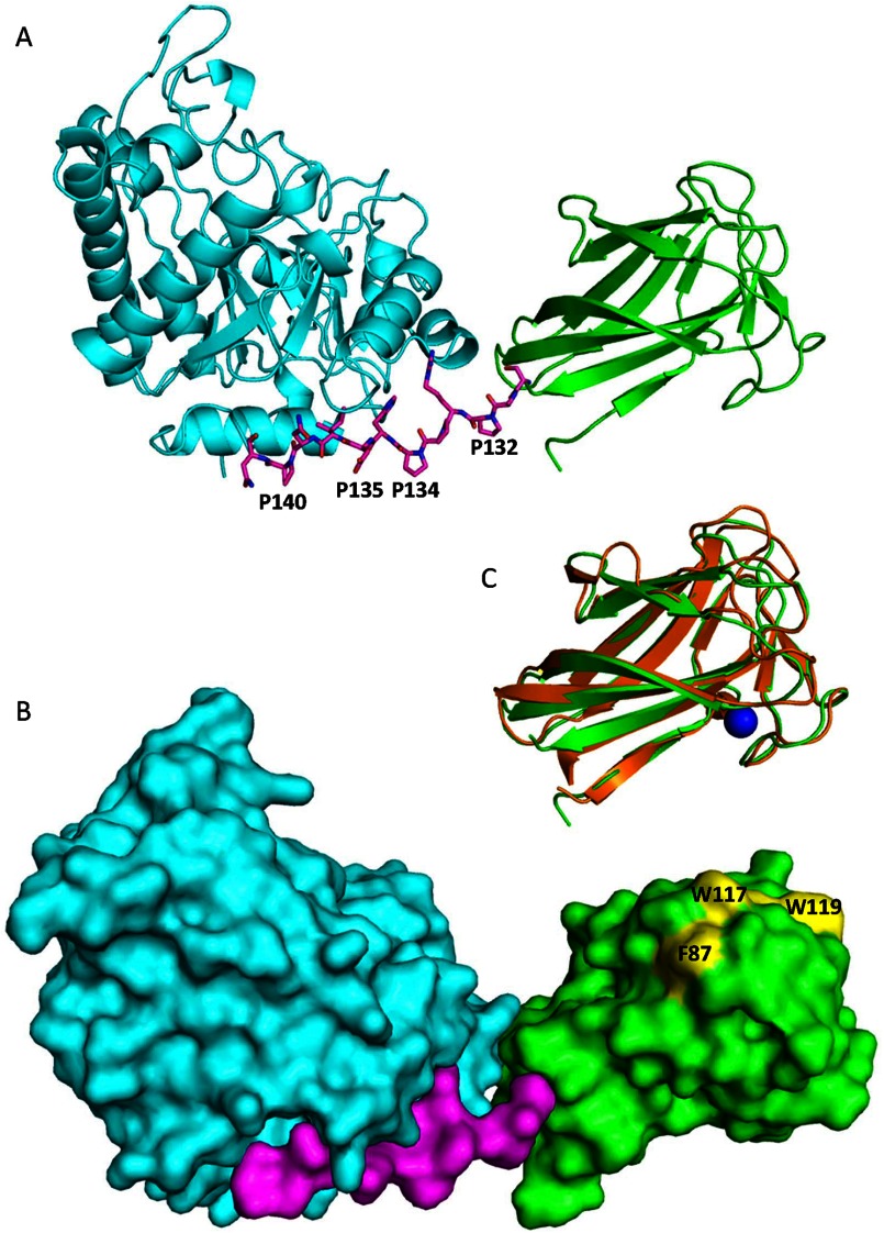
FIGURE 6.
Views of Modular architecture of PaMan26A. A, shown is a ribbon diagram of PaMan26A catalytic (blue) and CBM (green) domains. The proline-rich linker is shown in stick format. B, shown is a molecular surface representation of PaMan26A structure with the catalytic domain in blue, the PaCBM35 domain in green, and the linker in purple. The three aromatic residues present at the surface of the PaCBM35 domain are shown in yellow. C, shown is superposition of the PaCBM35 domain (green) and C. thermocellum CBM35 (orange). The calcium ion is represented by a blue sphere.

