Figure 1.
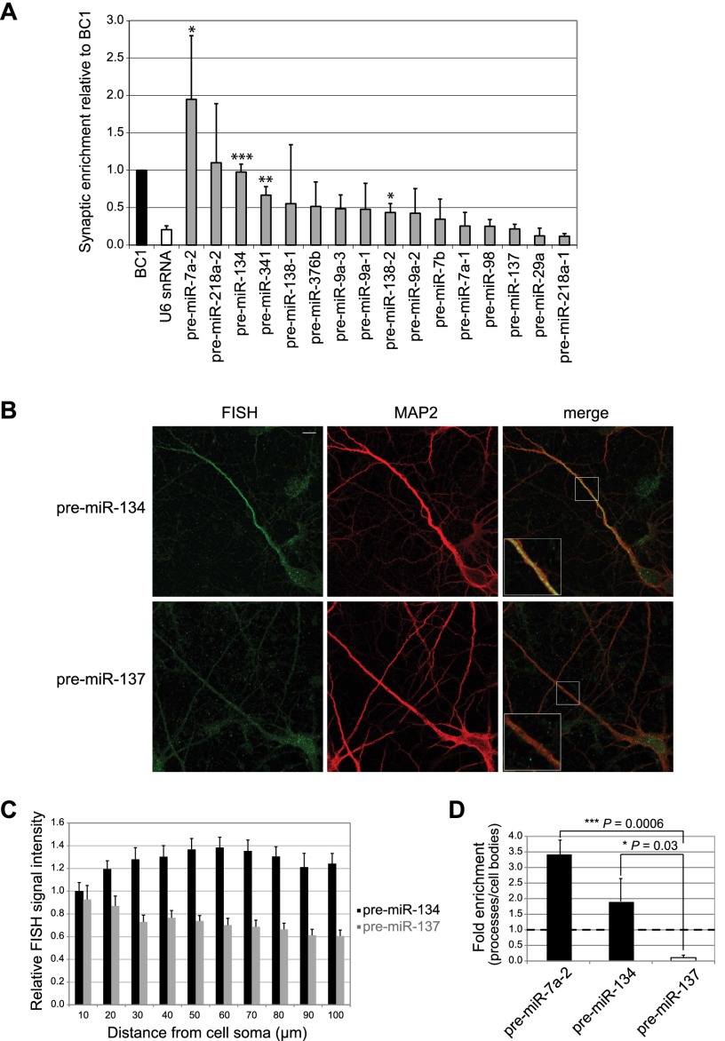
pre-miR-134 localizes to neuronal dendrites and synapses. (A) Levels of the indicated pre-miRNAs, BC1, and U6 snRNA in rat postnatal day 15 (P15) synaptosomes relative to whole forebrain measured by qRT–PCR (mean ± SD, n = 3). (*) P < 0.05; (**) P < 0.01; (***) P < 0.001). BC1 was set to one. (B, left) Representative images from FISH on BDNF-treated hippocampal neurons (7 DIV) using LNA probes directed against the terminal loop of either pre-miR-134 (top) or pre-miR-137 (bottom). (Middle) MAP2 immunostaining. (Right) Merge. Inserts at higher magnification illustrate the presence of pre-miR-134 puncta and the absence of pre-miR-137 puncta in distal dendrites. Bar, 10 μm. (C) Quantification of FISH analysis performed in B. Relative signal intensities of dendritic segments derived from 20 neurons of each condition ±SD. (D) Levels of indicated pre-miRNAs in the process compartment of hippocampal neurons relative to cell bodies measured by qRT–PCR (mean ± SD, n = 3).

