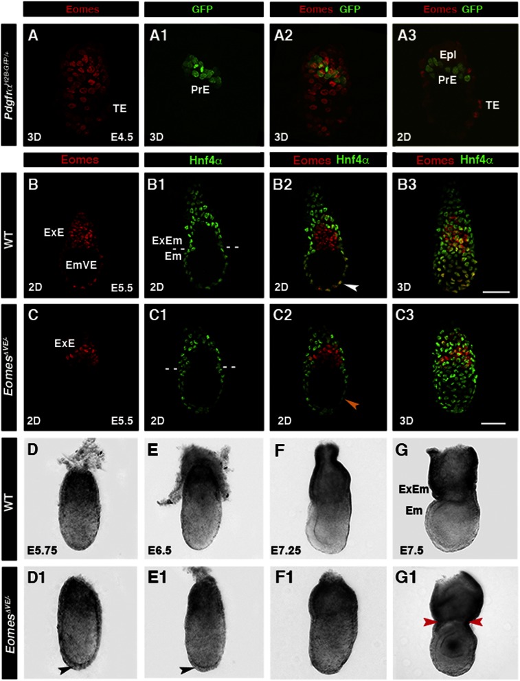
Figure 1.
Eomes is activated in the EmVE, and genetic ablation in the VE results in morphogenesis defects. (A–A3) E4.5 PdgfrαH2B-GFP embryo depicting nuclear-localized Eomes (red) in trophectoderm (TE) and absence in primitive endoderm (PrE). (B–B3) Colocalization of Eomes (red) and Hnf4α (green) in the EmVE (white arrowhead). (C–C3) Localization of Eomes and Hnf4α in E5.5 embryo with VE-specific Eomes inactivation (EomesΔVE/−). Note the lack of colocalization of Eomes and Hnf4α protein (C2; orange arrowhead). (D,D1) E5.75 EomesΔVE/− mutant embryos (D1) exhibit increased thickening of the VE at the distal tip (D1; black arrowhead) compared with wild-type (WT; D). (E,E1) At E6.5, the AVE has migrated anteriorly in wild type (E) but remains thickened distally in EomesΔVE/− mutants (E1; black arrowhead). At E7.25 (F,F1), EomesΔVE/− mutants display aberrant morphology (F1) compared with wild type (F), becoming exacerbated by E7.5 (G1). (G,G1) Note constriction at embryonic/extraembryonic junction (ExEM) (red arrowheads). (2D) Single optical section; (3D) projection of z-stack; (Em) embryonic; (Epi) epiblast; (ExE) extraembryonic ectoderm.

