
Figure 5. CD4+ T cell-specific deletion of IL4ra abolished neonatal RSV infection-induced immunopathophysiology upon reinfection.
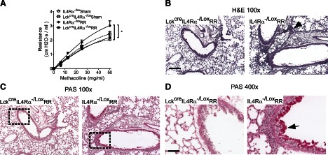
Th-specific, IL-4Rα-deficient (LckcreIL4Rα−/Lox-RR) and -competent mice (IL4Rα−/Lox-RR) were infected with RSV at 5 days old and reinfected at 4 weeks postprimary infection. Control mice of each genotype were treated with serum-free medium (LckcreIL4Rα−/Lox-sham and IL4Rα−/Lox-sham, respectively). Pulmonary function and lung histopathology were determined at 6 days postreinfection. (A) Airway resistance was measured at baseline (0 mg/ml MeCh) and increasing doses of intratracheally delivered MeCh to quantify airway hyper-reactivity. At baseline, airway resistance was not different between the groups; therefore, individual responses were normalized to their prospective baselines and normalized resistance plotted; n = 5–7; *P < 0.05. (B) H&E staining, demonstrating pulmonary inflammation. The arrow points to cellular infiltrates. Original scale bar = 50 μm. (C) PAS staining showing airway mucus. The arrow points to mucus staining. The right panels are enlargements of the rectangles in the left panels. Original scale bar = 200 μm.