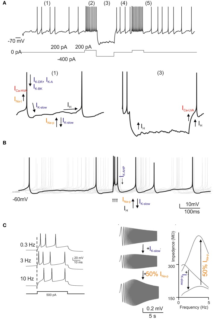
Figure 3.
Golgi cell ionic mechanisms. This is a reconstruction of the ionic mechanisms of the Golgi cell membrane obtained using computational models (Solinas et al., 2007a,b) based on previous electrophysiological analysis (Forti et al., 2006) and incorporated into a large-scale granular layer model network (Solinas et al., 2010). Transient Na current (INa − t); persistent Na current (INa − p); resurgent Na current (INa − r); high-voltage-activated Ca current (ICa − HVA); Ca-dependent K current of the BK-type (IK − BK); Ca-dependent K current of the SK-type (IK − AHP); delayed-rectifier K current (IKV); slow K current of the M-type (IK − slow); fast-inactivating K current of the A-type (IK − A); slow inward-rectifier H-current (Ih). In the different panels, the ionic channels involved are shown with arrows indicating their depolarizing or hyperpolarizing action. (A) Golgi cell responses like those reported in Figure 1A can be elicited by the model: (1) low-frequency pacemaking, (2) high-frequency spike discharge upon current injection, (3) sagging inward rectification, (4) post-inhibitory rebound, (5) phase resetting. (B) Golgi cell responses to bursts in the mossy fibers (arrows). After a burst, all responses are phase reset, generating an apparent “silent pause”. (C) Golgi cell responses to bursts in the mossy fibers repeated at different frequencies. Note that maximum responses are obtained around 6 Hz. The panels on the right show a “ZAP” protocol for investigating resonance. Resonance is determined by IK − slow and is amplified by INa − p.

