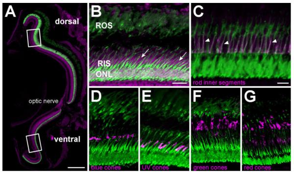
Figure 1.
NTR-EGFP expression is restricted to all rod photoreceptor cells in adult Tg(zop:nfsB-EGFP)nt19 fish. A: Confocal microscopy of TO-PRO-3 (magenta) counterstained retinal cryosections revealed fluorescent-labeled rod photoreceptors distributed throughout the entire ONL of 4–6-month-old Tg(zop:nfsB-EGFP)nt19 retinas. Rectangles represent the approximate 150 μm dorsal and ventral regions where PCNA-positive cells were quantified in metronidazole-treated fish. B: A higher magnification of the TO-PRO-3 counterstained retina revealed EGFP expression in rod nuclei in the ONL, as well as the RIS and ROS. EGFP is clearly not expressed in the cone cell nuclei adjacent to the RIS (B, arrows). C: All RIS labeled with the zs-4 marker (magenta) also expressed the transgene (arrowheads). D–G: EGFP-positive cells in the Tg(zop:nfsB-EGFP)nt19 retina clearly did not co-label with any of the cone opsins (magenta): blue (D), UV (E), green (F), and red (G). ROS, rod outer segments; RIS, rod inner segments; ONL, outer nuclear layer. Scale bar = 150 μm in A; 20 μm in B (applies to B,D–G); 10 μm in C.

