
Figure 10.
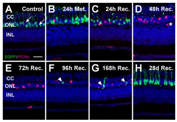
Proliferation response to ablation of a subset of rod photoreceptors. To examine regeneration following loss of a portion of rods in the dorsal metronidazole-treated Tg(zop:nfsB-EGFP)nt20 retina, proliferating cells were labeled with PCNA (magenta) and nuclei were stained with TO-PRO-3 (blue). A: Only a subset of the rods in the control retina expressed the nfsB-EGFP transgene, based on the many EGFP-negative rod cell nuclei in the ONL (arrows). B: EGFP- expressing rods became disorganized and ONL rod precursor proliferation was slightly increased after 24 hours of metronidazole treatment. C: By 24 hours of recovery, loss of EGFP-expressing rods and the presence of EGFP-positive pyknotic ONL nuclei confirmed the presence of rod cell death, while there was also an increase in the number of PCNA-positive nuclei in the ONL. D,E: Virtually all EGFP- expressing rods were absent by 48 (D) and 72 (E) hours of recovery, with even greater numbers of PCNA-expressing ONL nuclei, corresponding to committed rod precursor cells. A single PCNA-positive cell was observed in the INL at 48 hours of recovery, whose localization was not consistent with proliferating Müller glial cells (D, asterisk). F,G: Regenerated EGFP-positive rods appeared at 96 (F) and 168 (G) hours of recovery (arrowheads). H: After 28 days of recovery, EGFP expression in the ONL was similar to the control, indicating that new rod photoreceptors had fully regenerated. Notably, the ONL contained TO-PRO-3-labeled nuclei at every time point, confirming that rods lacking the transgene were not killed. Furthermore, cone cell nuclei, located apical to the ONL rod cell nuclei, were clearly discernable at all times in the cone cell layer. CC, cone cell layer; ONL, outer nuclear layer; INL, inner nuclear layer. Scale bar = 20 μm in A (applies to A–H).