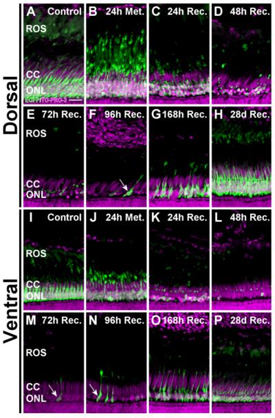
Figure 3.
Dorsal-ventral differences in photoreceptor loss following metronidazole treatment. Metronidazole-treated Tg(zop:nfsB- EGFP)nt19 retinal cryosections were stained with TO-PRO-3 and examined for loss of EGFP fluorescence. A,L: The ONLs of control retinas were comprised of healthy, EGFP-positive rod photoreceptors in the dorsal (A) and ventral (I) retinal regions. B,J: At the end of the 24-hour treatment, the ROS appeared to condense and retract basally, toward the ONL. C,K,D,E: Loss of EGFP-positive cells was apparent by 24 hours of recovery (C,K) and continued in the dorsal retina until virtually no EGFP-labeled nuclei were detected in the ONL after 72 hours of recovery (D,E). F,G: Newly regenerated, EGFP-expressing immature rods were observed at 96 (F) and 168 (G) hours of recovery (arrow). L: In the ventral retina, maximal cell loss was achieved by 48 hours of recovery. M–O: At 72–168 hours of recovery, EGFP-expressing immature rods were detected in the ventral retina (arrows). H,P: By 28 days of recovery, both the dorsal and ventral ONL had regenerated to a thickness comparable to the control. The NTR-EGFP-negative cone cell nuclei, located apical to the rod nuclei in the cone cell layer, were retained throughout this time course. ROS, rod outer segments; CC, cone cell layer; ONL, outer nuclear layer. Scale bar = 15 μm in A (applies to A–P).

