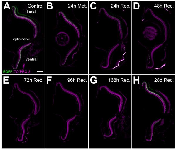
Figure 8.
Rod photoreceptor cell loss and regeneration following metronidazole treatment of Tg(zop:nfsB-EGFP)nt20 zebrafish. Tg(zop:nfsB- EGFP)nt20 zebrafish were treated with 10 mM metronidazole for 24 hours and transferred to a recovery tank. Eye specimens were collect from untreated controls, after 24 hours of metronidazole treatment, and after 24, 48, 72, 96, 168 hours, and 28 days of recovery. Death of NTR-EGFP expressing rod photoreceptors was assessed by loss of EGFP fluorescence. A: EGFP-positive cells were observed in the dorsal and ventral ONL of adult control retinas. B: After 24 hours of metronidazole treatment, there was a reduction and disorganization of the EGFP expression in the ONL. C,D–F: Cell loss was evident by 24 hours of recovery (C), with few EGFP-positive rods detected from 48 through 96 hours of recovery (D–F). G,H: Rods were beginning to regenerate by 96 and 168 hours of recovery (G) and the metronidazole-treated retinas were comparable to controls by 28 days of recovery (H). There was no significant difference in cell loss or regeneration between the dorsal and ventral regions. ONL, outer nuclear layer. Scale bar = 150 μm in A (applies to A–H).

