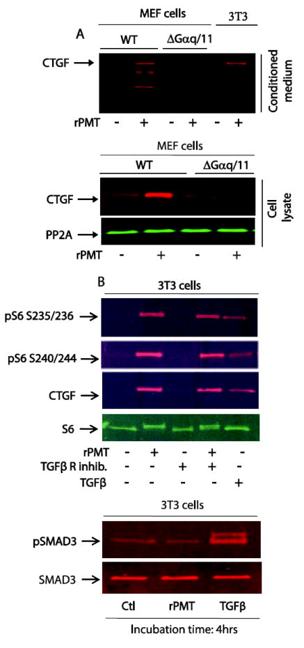
Figure 6. rPMT-induced CTGF protein is Gαq/11 dependent.

MEF wild type and MEF Gαq/11 deficient cells were grown in DMEM containing 10% of FBS till confluency. Confluent cells were switched to serum-free DMEM media for 48h and then treated with 100 ng/ml of rPMT for 24h. Control and rPMT treated cells were lysed in SDS PAGE loading buffer and equal amounts of proteins were separated on SDS polyacrylamide gel electrophoresis and transferred onto nitrocellulose membranes. Proteins in conditioned medium were also separated and transferred as described. CTGF protein levels were monitored by Western blot in both whole cell extract and conditioned medium. An antibody that recognizes protein phosphatase 2A was used for loading control (A). (B) 3T3 cells were serum-starved as above and treated with 50 ng/ml of rPMT or 20 ng/ml of TGFβ for 4 hours in the presence or absence of TGFβ receptor inhibitor. Cells were lysed and proteins were separated by SDS PAGE. Immunoblots were developed with antibodies that recognized unphosphorylated S6 (S6), phosphorylated S6 (pS6), CTGF, SMAD3 and phoaphorylated SMAD3 (pSMAD3).
