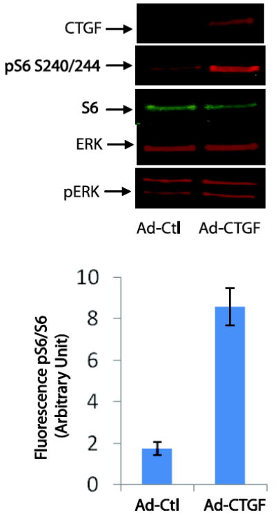
Figure 9. CTGF overexpression induces S6 phosphorylation.

Swiss 3T3 cells were grown in DMEM containing 10% of FBS till confluency. Confluent cells were switched to serum-free DMEM media and immediately transfected with Adenovirus ctl (Ad-Ctl) or adenovirus-CTGF (Ad-CTGF) for 24h. Cells were then harvested and lysed in SDS PAGE loading buffer. The same amount of protein were separated on SDS PAGE and transferred to nitrocellulose membranes. Proteins were probed using antibodies that recognize CTGF, ERK, pERK, S6 and pS6. The bar graph represents the mean +/- SEM from three independent experiments.
