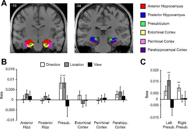
Figure 4.
Coding of spatial quantities in multivoxel patterns in the medial temporal lobe. A, Six anatomical regions were defined in the medial temporal lobes of each subject as described in Materials and Methods. The six regions from one subject are displayed on two coronal slices. B, Multivoxel patterns in presubiculum distinguished between directions and between locations. No other region showed coding of spatial quantities. C, Data for presubiculum, shown separately for each hemisphere, which suggests a difference in spatial coding across hemispheres. Left presubiculum distinguished between locations, whereas right presubiculum distinguished between directions. Presub, Presubiculum; Hipp, hippocampus; Parahipp, parahippocampal. **p < 0.01; *p < 0.05. Error bars indicate mean ± SEM.

