
Figure 7.
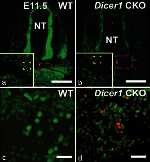
EdU (green) and ApopTag (red) staining in coronal sections of E11.5 Dicer1 CKO. a, c. WT. b, d. Dicer1 CKO. High magnification of dotted boxes shown in white inset box. NT: neural tube. Bar = 500 μm (a, b); 20μm (C, D)
Official websites use .gov
A
.gov website belongs to an official
government organization in the United States.
Secure .gov websites use HTTPS
A lock (
) or https:// means you've safely
connected to the .gov website. Share sensitive
information only on official, secure websites.
EdU (green) and ApopTag (red) staining in coronal sections of E11.5 Dicer1 CKO. a, c. WT. b, d. Dicer1 CKO. High magnification of dotted boxes shown in white inset box. NT: neural tube. Bar = 500 μm (a, b); 20μm (C, D)