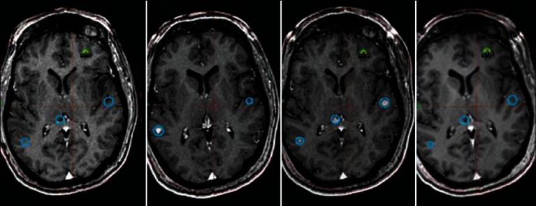
Figure 4.

Sixty-six-year-old male with metastatic renal cell cancer. Thirty-three lesions have been treated over two and one-half years. MRI shows representative images of the same anatomical location over time with the oldest scans on the left and newest on the right. The blue circles represent previously treated lesions and the yellow/green represent lesions most recently treated
