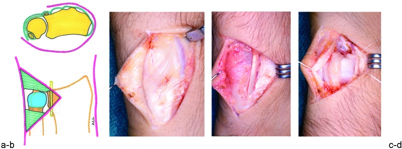
Fig. 1 .

(a) (Top) Cross-section anatomy of the DRUJ. The forearm is in pronation, and the ulnar head is covered by the DRUJ capsule and the extensor retinaculum. The fifth extensor compartment stabilizes the extensor digiti minimi tendon at the most ulnar border of the distal radius. The extensor carpi ulnaris (ECU) tendon is stabilized by its own sheath, separate from the extensor retinaculum, lateral to the styloid process of the ulna. (Bottom) Ulnar head after the extensor retinaculum has been ulnarly reflected and the dorsal radioulnar capsule removed. (b) Exposure of the extensor retinaculum. (c) Division of the fifth extensor compartment allows for identification and radial displacement of the extensor digiti minimi tendon. (d) Longitudinal division of the capsule exposes the head of the ulna. Joint synovitis, when present, should be removed as exposure will improve.
