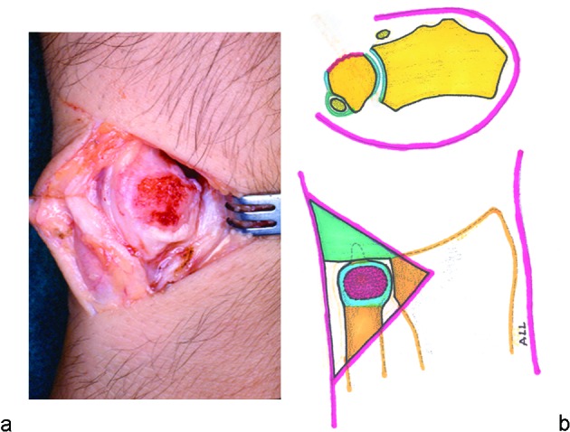
Fig. 2 .

(a) With the forearm in pronation, the joint cartilage and subchondral bone of the ulnar head facing the surgeon are removed, leaving a slightly convex surface of cancellous bone. (b) The ulnar head is perforated using a 3.2-mm drill bit, with the entrance at the center of the denuded ulnar head, which exits anteriorly to the styloid process and ECU sheath. The direction of the drill should be perpendicular to the long axes of the radius and ulna.
