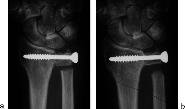
Fig. 7 .

(a) Radiograph of the wrist showing the head of the ulna compressed against the radius with a malleolar screw, with a very small segment of ulna removed. (b) With the passing of time, the bone defect will get larger from resorbtion of the bone ends, mainly the proximal ulna stump.
