
Figure 1. nkx2.5-expressing mesoderm guides pouch epithelial transitions.
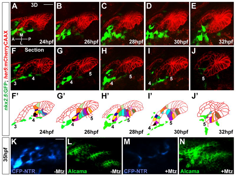
(A-E) 3D projections captured from time-lapse recording of wild-type pouch development (See Movie S2) show intimate interactions between her5:mCherryCAAX-positive endodermal epithelia (red) and nkx2.5:GFP-positive mesoderm (green). Anterior-posterior (A-P) and medial-lateral (M-L) axes are shown.
(F-J) Representative sections from the same time-lapse recording (see Movie S2) show various stages of development of pouches 3-5. In the schematics (F’-J’), the tracking of individually color-coded pouch cells highlights cell rearrangements. Cells that we could not track through the entire recording were left uncolored.
(K-N) CFP fluorescence (blue) and Alcama immunohistochemistry (green) show that injection of 5 nl of 5 mM Mtz into nkx2.5:Gal4VP16; UAS:CFP-NTR embryos results in reductions of CFP-NTR-expressing mesoderm and disorganized pouch endoderm (M and N) compared to un-injected siblings (K and L). Scale bars = 20 μM.