
Figure 3. Roles of Wnt signaling components in pouch and CB cartilage development.
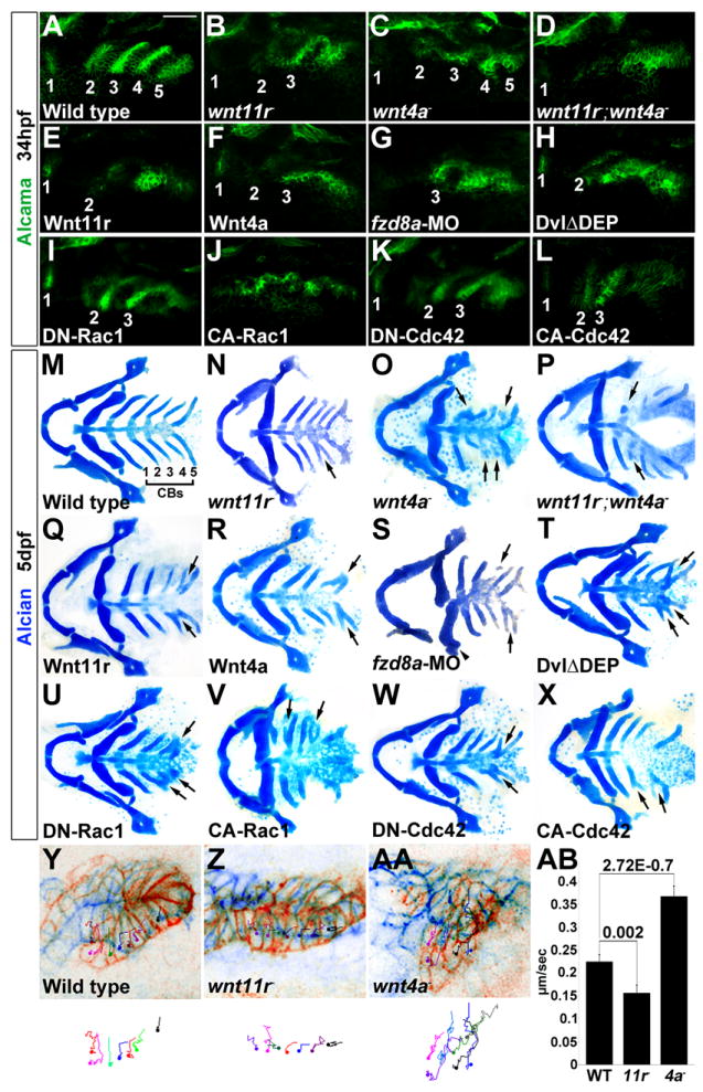
(A-L) Alcama immunohistochemistry (green) shows defects in pouches in mutant, fzd8a-MO, and transgenic embryos. Identifiable pouches are numbered. UAS transgenic embryos (capitalized) were doubly positive for nkx2.3:Gal4VP16. Scale bar = 40 μM.
(M-X) Whole mount views of dissected facial cartilages. Arrows indicated fused or abnormal CB cartilages. Arrowhead indicates abnormal hyoid cartilage in fzd8a-MO embryos.
(Y-AA) Superimposition of initial (blue) and final (red) still images from time-lapse recordings of fifth pouch development in wild-type, wnt11r-/-, and wnt4a-/- embryos. Colored lines indicate cell tracks (shown also below merged images), with filled circles denoting final positions.
(AB) Average pouch cell speed in wild types and mutants. Data represent mean ± SEM and p-values are shown for each comparison.
See also Figures S1 and S2.