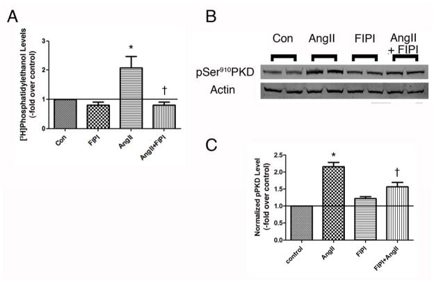
Figure 4. FIPI inhibits AngII-induced PLD and PKD activation in the human adrenocortical carcinoma cell line, HAC15.
(A) [3H]Oleate-prelabeled HAC15 cells were pretreated for 20–30 minutes with or without 750 nM FIPI (or the DMSO vehicle) prior to incubation in the presence or absence of 10nM AngII for 30 minutes. Cell lipids were extracted using chloroform-methanol and analyzed by thin-layer chromatography. Results represent the means ± SEM of 3 separate experiments performed in duplicate; *p<0.05 versus the control, †p<0.05 versus AngII alone. (B) HAC15 cells were pretreated for 30 minutes with or without 750 nM FIPI (or the DMSO vehicle) prior to incubation in the presence or absence of 10nM AngII for 30 minutes. Autophosphorylation of PKD at serine910 was determined by western analysis, with a representative blot of duplicate samples shown in panel B. (C) Phosphoserine910 levels were normalized to total PKD levels and expressed as fold relative to control. Values represent the means ± SEM of 5 separate experiments performed in duplicate; *p<0.05 versus the control, †p<0.05 versus AngII alone.

