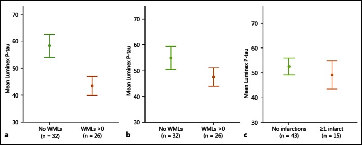
Fig. 1.
CSF levels of P-tau in prodromal AD patients with or without WMLs. The P-tau levels are significantly lower in patients with WMLs in the parietal lobes (a), but not in patients with WMLs in the frontal lobes (b) or in patients with infarctions in the basal ganglia (c). Error bars represent ± 1 SE.

