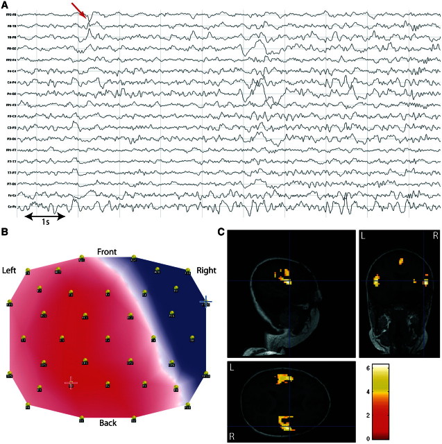
Figure 8.
Discordant results in medial/polar temporal lobe epilepsy. Patient 19 with right temporal epilepsy, dysplasia of right uncus. (A) Long-term EEG. Red arrow = representative spikes used to build the epileptic map; (B) epileptic map derived from long-term EEG, blue/red cross indicates maximum negativity/positivity; (C) topography-related BOLD changes (P < 0.001, uncorrected for display but the bilateral opercular activations survived FWE correction, P < 0.05) co-registered with postoperative MRI (right anterior temporal lobectomy).

