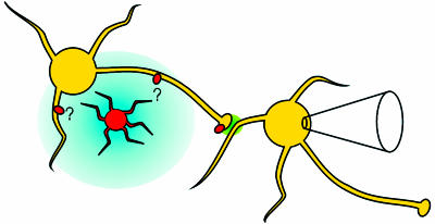
Fig. 1.
A model showing the circuit involved in the experiments of Liu et al. (9). An astrocyte (orange cell) is loaded with caged calcium. Upon photolysis, the astrocyte releases glutamate (blue), which spreads to activate GluR5-containing kainate receptors (red ovals) on nearby hippocampal interneurons (yellow cells). The activation of kainate receptors leads to increased firing of the interneuron, causing enhanced spontaneous GABA release (green). This enhanced release is detected as an increase in the frequency of sIPSCs on a nearby interneuron, monitored by patch–clamp recording. The range of glutamate after release from the astrocyte is unknown, as is the subcellular location of the interneuronal kainate receptors that sense it.

