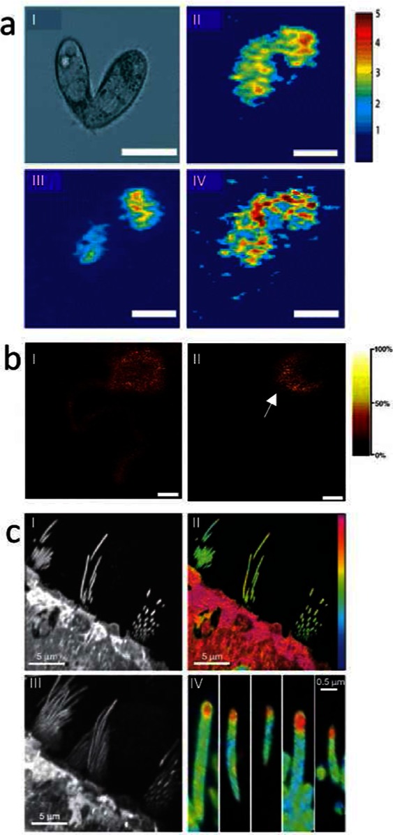
Figure 2.

Fundamental neuroscience research imaging mass spectrometry. (a) SIMS imaging reveals distribution of lipid species that are coupled to membrane curvature in mating cells.69 (I) Differential interference contrast microscopy image of a mating cell pair. (II–IV) SIMS ion images of a triturated pair of mating T. thermophila for different lipid species, including C5H9 (I), PC (II), and 2-AEP (IV) (the intensity in the 2-AEP image has been multiplied by factor 3) (scale bar = 25 um). (b) Application of SIMS for imaging single neurons allows subcellular localization of vitamin E. (Reproduced with permission from ref (65). Copyright 2005 American Chemical Society.) (I) Neuronal lipid distribution illustrated by the choline fragment trimethylethenamine (m/z 86), derived from sphingosine and phosphatidylcholin (PC), showing homogeneous distribution throughout soma and neurite. (II) Interestingly, vitamin E was found to distribute to the soma–neurite junction, suggesting an important role in neuronal communication (II) (scale bar = 100 μm). (c) Multi isotope imaging MS was employed at 30 nm resolution to demonstrate protein turnover in hair cells.80 (I) SIMS ionimage of protein fragment CN (m/z 26) of utricle; day 56 and ration CN15/CN14 (m 27/m 26) showing low incorporation in stereocilia. (III) Projection of a three-dimensional stack of image (I). (IV) CN15/CN14 Ratio image from (III) reveals high protein turnover to occur at a high rate at the tip of hair cells.
