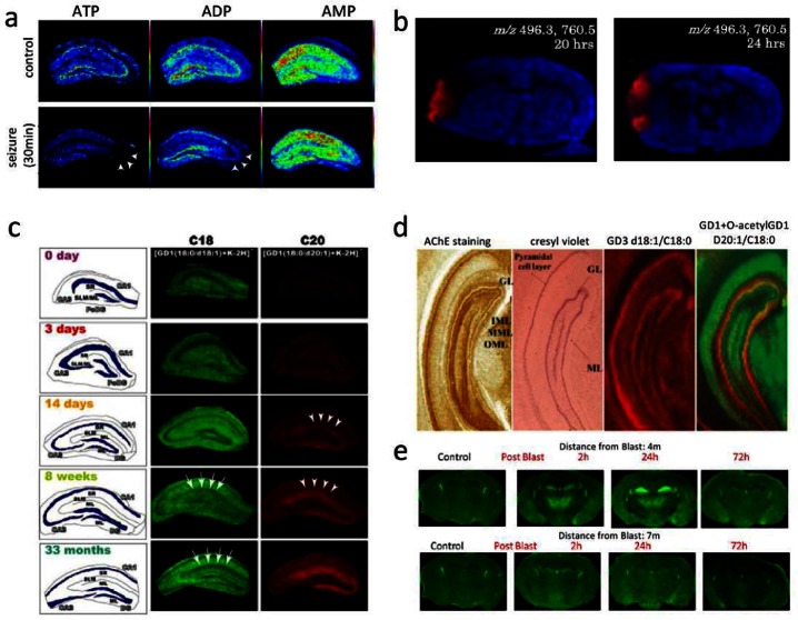
Figure 3.
Imaging mass spectrometry in clinical and molecular neuroscience for low molecular weight compounds and lipids. (a) Investigation of energy metabolism in hippocampus formation reveals selective ATP/ADP turnover in CA3 following induced seizure (ATP/ADP, arrows).83 (b) IMS based evaluation of ischemia shows regional selective increase of LPC (m/z 496) following injury 20 and 24 h.85 (c) Application of MALDI imaging for mapping ganglioside species reveals aging dependent regional increase of C20 gangliosides in the molecular layer (ML) of the dentate gyrus (DG) (14 days and 18 weeks, C20 arrows).91 (d) Using a similar approach, IMS revealed characteristic distributions of gangliosides in the hippocampus based on their ceramide core. In addition, in situ characterization of unknown glycolipid modifications as demonstrated by the discovery of O-acetylation (right overlay image). (Reproduced with permission from ref (92). Copyright 2011 American Chemical Society.) (e) IMS based ganglioside imaging reveals selective C20-ganglioside increase in, e.g., the hippocampal formation following blast induced mild TBI (2 and 24 h past blast, at 4 m but not 7 m distance). (Reproduced with permission from ref (93). Copyright 2013 American Chemical Society.)

