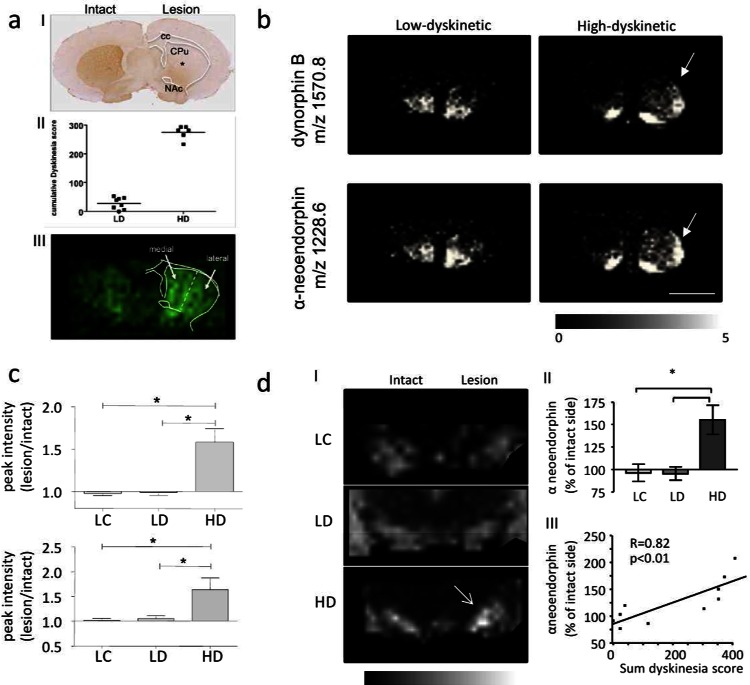
Figure 4.
Imaging mass spectrometry of neuropeptides in L-DOPA induced dyskinesia. (a) (I) Unilateral 6-OHDA injection leads to dopamine depletion (as illustrated by TH immunostaining*); (II) L-DOPA therapy results in two distinct groups with low and high-dyskinesia; (III) MALDI IMS and assignment of ROI and spectra extraction. (b,c) Similarly, dynorphin peptides were found significantly increased in the dorso-lateral striatum of high- (HD) compared to low -dyskinetic (LD) animals and lesion controls (LC). (For panels (a)–(c), this research was originally published in Molecular Cellular Proteomics. Hanrieder et al. Mol. Cell Proteomics2011; M111.009308. Copyright the American Society for Biochemistry and Molecular Biology.32) (d) (I,II) MALDI IMS reveals significant increase of dynorphin peptide (here alpha neoendorphin) in the substantia nigra (SN). (III) Dynorphin peptide intensity correlated positively with dyskinesia severity.95

