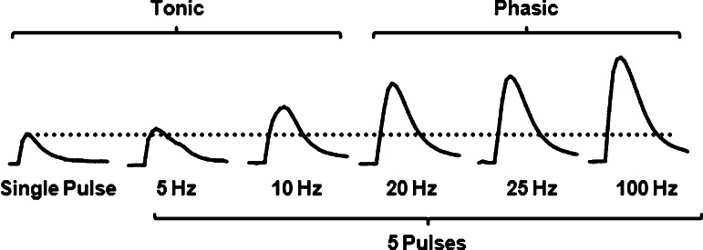
Figure 2.
Representative dopamine traces highlighting the frequency response in the magnitude of dopamine in the nucleus accumbens shell. Electrical stimulations during voltammetric recording of evoked DA in brain slices may vary in pulse number, current amplitude, and frequency to model both tonic and phasic firing of DA neurons that occur in vivo. One common approach is to evaluate evoked DA release to single pulse and multiple pulses (e.g., 5) across frequencies that range from 5 to 100 Hz, with 20 Hz as a general tipping point in the shift from tonic to phasic signaling of DA neurons. Frequency response is more robust in the ventromedial striatum (i.e., nucleus accumbens shell) as shown here compared to the dorsolateral striatum, although the phasic frequency that elicits the highest peak amplitude can vary. Ratios can be calculated that compare the peak-height of the phasic signals to either the peak height of single pulse signal (dotted line), or the peak-height of a multiple pulse tonic signal.

