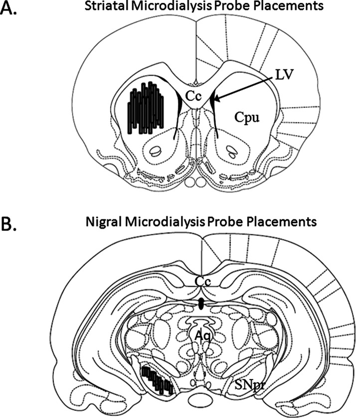
Figure 3.
Microdialysis Probe Placements. Schematic representations of coronal rat brain sections taken from Paxinos and Watson (1998). Shaded cylinders depict the distribution of (A) striatal (bregma 1.20 mm) and (B) nigral (bregma −5.30 mm) probe sites in all rats used in Experiment 2 (N = 26). Relevant anatomical structures: Aq, aqueduct (Sylvius); Cc, corpus callosum; Cpu, caudate putamen; LV, lateral ventricle; SNpr, substantia nigra pars reticulata.

