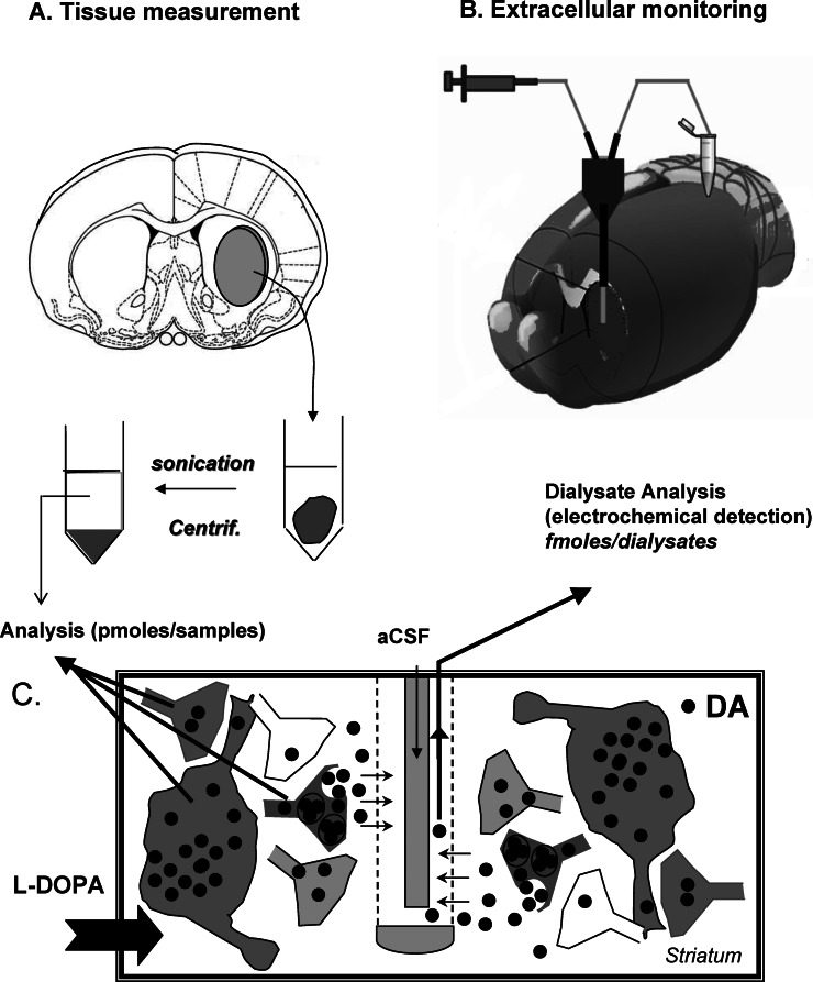
Figure 1.
Contribution of intracerebral microdialysis to the mechanism of action of L-DOPA toward dopamine responses. (A) Tissue measurement in response to L-DOPA administration in rodents was originally used. Immediately after the animal was sacrificed, the brain region of interest (here the striatum) was removed, placed in an acid medium, sonicated, and centrifuged. Monoamine tissue concentrations from the supernatant were quantified by high pressure liquid chromatography coupled to electrochemical detection (HPLC-ED). (B) Extracellular monitoring using intracerebral microdialysis is performed using microdialysis probes inserted in the brain region of interest (here the striatum of a living animal). The continuous flow rate of the artificial cerebrospinal fluid (aCSF) permits to collect samples at regular intervals. The dialysates are often analyzed with HPLC-ED due to the high sensitivity of this approach toward monoamines. (C) The panel illustrates the different origin of the DA signals analyzed with tissue measurement and intracerebral microdialysis after L-DOPA. L-DOPA will virtually enter all cells and/or terminals and, depending on the presence of L-DOPA decarboxylase, will be converted in DA in several loci. The magnitude of the DA signal is often large (picomoles) due to the contribution of multiple cellular systems. Using microdialysis probes, only DA reaching the extracellular space can be taken up by the probe. The contribution of cells to the DA signals, often low in magnitude (some femtomoles), is restricted to cells capable of releasing DA, e.g., the 5-HT neurons in DA-denervated rats.

