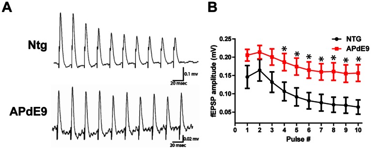
Figure 2. Impaired STP in the DG of APdE9 mice.

(A). Representative field potential responses recorded extracellularly in NTG mouse tissue. 40 Hz 10 pulse stimulation evoked depressive (decreasing amplitude) responses in the NTG animals. APdE9 mice showed sustained field potential responses with little decrease in the amplitude throughout the stimulation train. (B) Average and S.E.M. for APdE9and NTG fEPSP amplitudes during the 40 Hz stimulation. Note significant differences for the fEPSP amplitudes produced by pulse stimulations 4–10 (pulse to pulse comparison, unpaired t-test, p<0.05).
