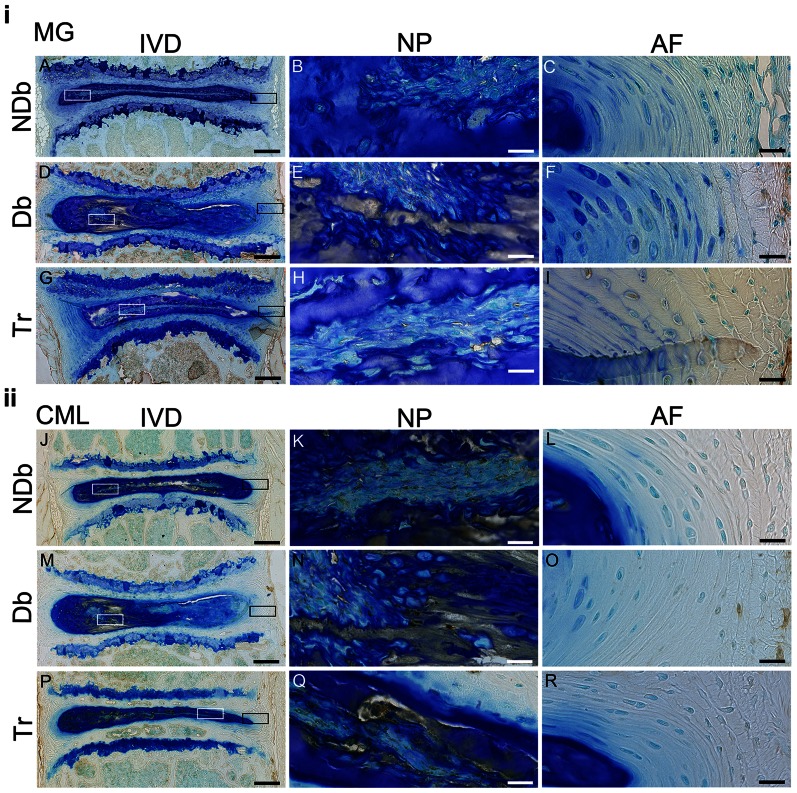
Figure 5. Diabetes causes AGE accumulation in IVD and vertebrae which is partially mitigated after treatment.
Representative images of immunohistochemistry for MG (top, A–I) and CML (bottom, J–R) of NDb, Db and Tr IVDs (left: 5× magnification); boxes mark 40× magnified area of NP (white box; MG = B,E,H and CML = K,N,Q) and AF (black box; MG = C,F,I and CML = L,O,R). Scale-bars: left (IVD) = 200 µm; right (NP+AF) = 20 µm.

