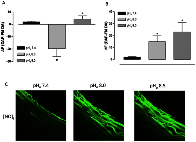
Figure 3. Effect of extracellular alkalinization on [NO]i of rat aorta cross sections assessed using a confocal scanning laser microscope.
A) Fluorescence intensity for endothelial layer. B) Fluorescence intensity for muscular layer. C) Representative confocal photomicrograph of one aorta cross section.. Aorta cross sections were loaded with DAF-FM DA (5 µM) and analyzed by confocal microscopy. Aorta cross sections were stimulated at 3rd and 9th min with Hanks alkalinized solution pH 8.0 and 8.5, respectively. NaOH was used to change the pH of Hanks solution; Hanks solution pH 7.4 served as control. Fluorescence intensity was measured before the stimulus (F) and at 6th min after the stimulus for each pHo value (FpH). Results are reported as ΔF = FpH – F. All values are means ± SEM (n = 7). One-way ANOVA, Bonferroni's post-test, # p<0.001 versus control, * p<0.05 versus control.

